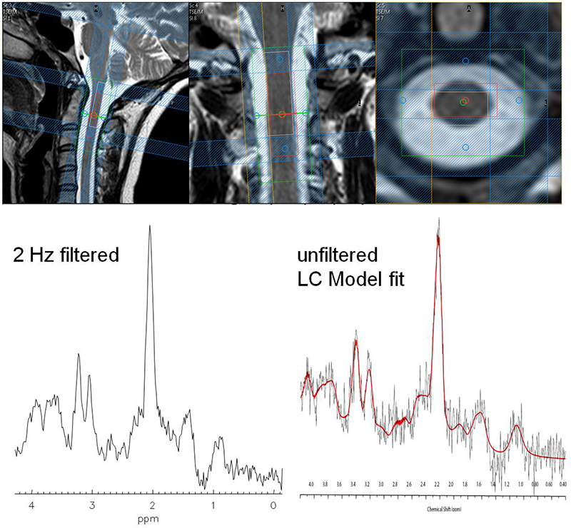
Spinal Cord MRS
Magnetic resonance spectroscopy (MRS) provides information about the biochemical processes of neuronal tissue that is complementary to conventional MRI investigations and is therefore a valuable tool for differential diagnostics of various neuropathologies. However, due to technical challenges MRS has been rarely applied to the spinal cord and was mainly restricted to the very upper part of the cervical spine.
Several malfunction sources could be identified besides the known difficulties in MRS, namely fluctuating B0 fields caused by respiratory motion, artifacts due to pulsatile flow of the cerebrospinal fluid (CSF) and patient movement, pronounced B0 inhomogeneities due to susceptibility differences between vertebral bodies and the nerve itself, cerebrospinal fluid (CSF) as well as muscle or lipid rich tissue outside the area of interest, and the low SNR achievable in the finite size of the spinal cord.
In this project methods shall be developed that improve the quality of spinal cord spectra in the cervical, thoracic and lumbar regions to make spinal cord MRS feasible for clinical diagnostics and physiological studies. Therefore, aspects should be investigated to realize decreased measurement time, optimization of the spectral quality, robustness, and significance of the measurement. More specifically, localization has to be robust and consistent for all metabolites of interest, water and lipid suppression as well as shimming reliable and reproducible and receive coils SNR optimized. Patient movement might be overcome by volume tracking, while ECG triggering with optimized trigger delays can minimize CSF flow artefacts. Fluctuations in the B0 field due to respiratory motion might be minimized of respiratory gating or dynamic shimming is used.
Publications:
- Henning A, Schär M, Kollias SS, Boesiger P, Dydak U Quantitative magnetic resonance spectroscopy in the entire human cervical spinal cord and beyond at 3T. Magn Reson Med 59(6), 1250-1258, 2008
Contact
No database information available

