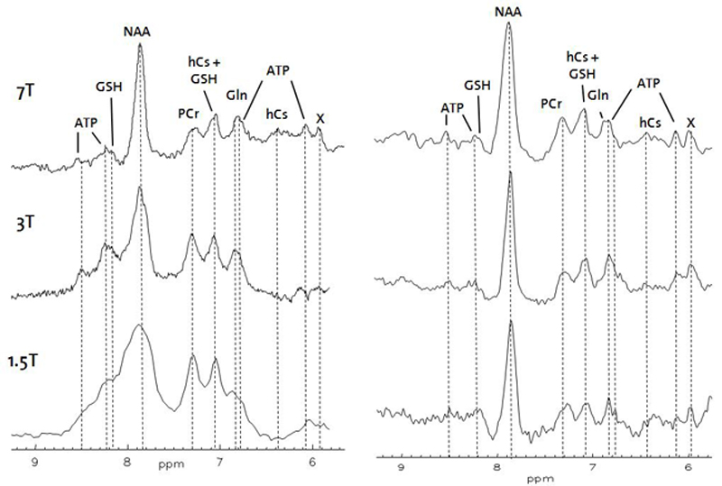
Downfield MRS
Profiting from SNR gain as well as increased spectral resolution, detection and quantification of an increasing number of metabolites becomes feasible at high (3T) and ultra-high field (7T) strength. While these advantages have been exploited for the upfield part of the human cerebral 1H spectrum, the downfield part (5-10ppm) where protons bound to carbon atoms in aromatic compounds, as well as amide protons can be observed, is still poorly characterized. Additional metabolites in comparison to the upfield spectrum such as ATP and homocarnosine (hCs) should be detectable; the amide protons of the NH2 group in glutamine (Gln) should allow its unambiguous distinction from glutamate. Potentially, additional resonances visible in the downfield part as well as changes in the chemical exchange rates of amide protons and the chemical shift of the imidazole ring resonances of hCs might become important disease markers. Prominent examples are the accumulation of phenylalanine in neuronal tissue of patients suffering from phenylketonuria or of Gln in hyperammonemia.
In this project, the complimentary information content of downfield spectra shall be assessed at 7T in order to improve spatial specificity and spectral resolution. To that ultra-high field related problems have to be overcome such as shortened T2 relaxation times and chemical shift displacement artefact, B1 and B0 inhomogeneity and artifacts such as gradient modulation sidebands have to be excluded.
To assist and validate assignments of downfield resonances 7T downfield MR spectra will be compared to corresponding spectra recorded at lower field strength and characterized regarding relaxation behaviour and in view of chemical exchange with water. Additional insight might be provided by pH titration series in phantoms and nutrition or infusion studies that might lead to an increase of the intensity of specific downfield resonance lines.
Publications:
- Henning A, Fuchs A, Boesch C, Boesiger P, Kreis R. Downfield spectra at ultra-high field. Proc. Intl. Soc. Mag. Reson. Med. 16 (2008) 777.
Contact
No database information available