Neuronal current DWI
Detection of neuronal currents (alpha waves) by EEG-MRI
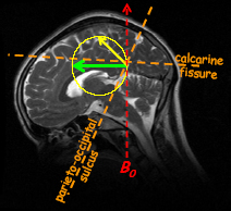
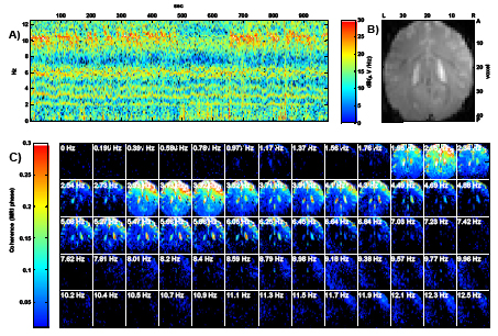
It has been suggested recently that the influence of the neuro-magnetic field should make electrical brain activity directly detectable by MRI. To test this hypothesis, we performed combined EEG-MRI experiments which aim to localize the neuronal current sources of alpha waves (8‑12Hz), one of the most prominent EEG phenomena in humans. A detailed analysis of cross-spectral coherence between simultaneously recorded EEG and MRI time series revealed no sign of alpha waves. Instead the EEG‑MRI approach was found to be hampered by artefacts due to cardiac pulsation, which extend into the frequency band of alpha waves. Separate brain displacement mapping experiments confirmed that not only the EEG but also the MRI signal is confounded by harmonics of the cardiac frequency even at 10Hz and beyond. This well known ballistocardiogram artefact cannot be avoided or eliminated entirely by available signal processing techniques. Therefore we must conclude that current EEG‑MRI methodology based on correlation analysis lacks not only the sensitivity but also the specificity required for the reliable detection of alpha waves.


References
- H. Mandelkow, P. Halder, D. Brandeis, M. Soellinger, N. de Zanche, R. Luechinger, and P. Boesiger, Heart beats brain: The problem of detecting alpha waves by neuronal current imaging in joint EEG-MRI experiments. Neuroimage, 2007. 37(1): p. 149-163.
- H. Mandelkow, P. Halder, P. Boesiger, and D. Brandeis, Synchronization facilitates removal of MRI artefacts from concurrent EEG recordings and increases usable bandwidth. NeuroImage, 2006. 32(3): p. 1120-1126.