DTI fiber verification
Verification of diffusion-weighted imaging and nerve fiber tracking
Diffusion tensor imaging (DTI) is a magnetic resonance imaging technique that enables the measurement of restricted water diffusion in the human brain. DTI provides quantitative measures which reflect the underlying tissue properties and exhibit markers in pathological states.
However, DTI data is fairly difficult to interpret in biological tissue as many factors affect the diffusion process and the MR signal. In addition to scalar diffusion parameters, the recently developed technique fiber tracking shows great promise for investigating the architecture of brain white matter. Presently, it is the only technique that enables in-vivo investigation of nerve fibers.
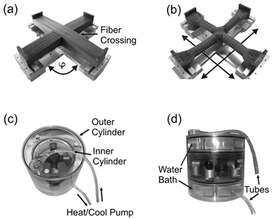
Therefore, verification of diffusion parameters and derived virtual axonal projections is an important ongoing research topic. Methods will be developed that enable in-vivo as well as ex-vivo investigation of results derived from diffusion-weighted imaging techniques.

References
- P. Staempfli, C. Reischauer, T. Jaermann, A. Valavanis, S. Kollias, and P. Boesiger, Combining fMRI and DTI: a framework for exploring the limits of fMRI-guided DTI fiber tracking and for verifying DTI-based fiber tractography results. Neuroimage, 2008. 39(1): p. 119-126.